Projects
home


Mutants

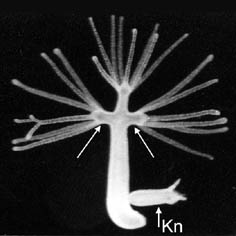
The mutant strain multi-headed-1 (mh-1) is a sexual inbreed generation of the wildtype (wt 105) of Hydra magnipapillata. It is able to produce several ectopic heads along its body axis (see figure, arrows).

By producing chimeras of various cell types we could show that the cause for the ability to form extra heads is located in the ectodermal epithelial cells of mh-1. The interstitial cells are not involved in this process. 
Mh-1 shows a kind of morphogenesis anusual for Hydra. Young mh-1 buds are morphological not distingiushable from wildtype buds. It takes nine days after separation from the parent animal untill the animal is able to form a secondary head. The change in the pattern forming system is in our opinion coupled with an increase of positional value. It increases. On the assumption that pattern formation is hierarchically organised, the increase of the positinal value leads to an activation of a secondary system which controls and determines the formation of ectopic headstructures.
We found that the head inhibitory acting activity (perhaps morphogenes) that may represent the primary system, is decreased in double-headed mh-1 compared to mh-1 with normal morphology. This difference in activity in double headed mh-1 shows that the properties of the tissue in this animals have changed. The bud development is not influenced. 
We were able to show that phosphorylation is involved in the control of pattern formation in mh-1. A single pulse treatment with the serine/threonine protein phosphatase inhibitor cantharidin leads to the formation of ectopic heads in young mh-1 buds. There are some links that tyrosin protein kinases are also involved in the pattern forming system in mh-1. We are now looking for the important phosphorylated / dephosphorylated proteins. Furthermore we are interested in identifying genes in mh-1 that are important for phosphorylation processes involved in development in other organisms.
 

S. Zeretzke and S. Berking (1996) Analysis of a hydra mutant which produces extra heads along the body axis. Int. J. Dev. Biol. Suppl.1:271S

S. Zeretzke and S. Berking (2001) Pattern regulation of a Hydra strain which produces additional heads along the body axis. Int. J. Dev. Biol. 45: 431-439 (2001) (PDF_File)

S. Zeretzke and S. Berking (2002)  In the multiheaded strain (mh-1) of Hydra magnipapillata the ectodermal cells are
       responsible for the formation of additional heads and the endodermal epithelial cells for the reduced ability to regenerate a foot.
       Develop. Growth Differ. 44: 85-93 


home
Budding of polyps
In Hydra sp. (Hydrozoa) and in Cassiopea sp. (Scyphozoa) a bud develops at the gastral region of the parent animal. The tissue of the bud does not originate from a meristem but from the gastral tissue of the parent animal. An important difference to note is that in hydrozoa the tip of the bud develops into the head of the polyp while in (many) scyphozoa the tip develops into the opposite part, namely the foot. Therefore, the central question is whether the systems which are controlling pattern formation in these animals are fundamentally different. 

A summary of the latest experimental results and of the disscusion about models of control mechanisms of bud development in hydra can be found under Berking, 1998. There is a recent paper addressing bud development in Cassiopea sp. We offer a theoretical model of pattern formation which allows to understand bud development in hydrozoa and scyphozoa. 

Publications :

Berking, S. (1977). Bud formation in Hydra: Inhibition by an endogenous morphogen. Roux's Arch. Dev. Biol. 181, 215-225.
Berking, S. (1979). Analysis of head and foot formation in Hydra by means of an endogenous inhibitor. Roux's Arch. Dev. Biol. 186, 189-210.
Berking, S. (1980). Commitment of stem cells to nerve cells and migration of nerve cell precursors in preparatory bud development in Hydra. J. Embryol. exp. Morph. 60, 373-387.
Berking, S. (1986). Transmethylation and control of pattern formation in Hydrozoa. Differentiation 32, 10-16.
Berking, S. (1998). Hydrozoa metamorphosis and pattern formation. Current Topics in Developmental Biology 38: 81-131
Berking, S., and Gierer, A. (1977). Analysis of early stages of budding in Hydra by means of an endogenous inhibitor. Roux's Arch. Dev. Biol. 182, 117-129.
Berking, S., and Schüle, T. (1987). Ammonia induces metamorphosis of the oral half of buds into polyp heads in the scyphozoan Cassiopea. Roux's Arch. Dev. Biol. 196, 388-390.

Kehls, N.E., K. Herrmann and S. Berking (1999) The protein phosphatase inhibitor cantharidin induces head and foot formation in buds of Cassiopea andromeda (Rhizostomae, Scyphozoa) Int. J. Dev. Biol. 43:51-58
ABSTRACT The polyps of Cassiopea andromeda produce spindle shaped, freely swimming buds which do not develop a head (a mouth opening surrounded by tentacles) and a foot (a sticky plate at the opposite end) until settlement to a suited substrate. The buds, therewith, look very similar to the planula larvae produced in sexual reproduction. With respect to both, buds and planulae, several peptides and the phorbolester TPA have been found to induce the transformation into a polyp. Here it is shown that cantharidin, a serine/threonine protein phosphatase inhibitor, induces head and foot formation in buds very efficiently in a 30 minutes treatment, the shortest yet known efficient treatment. Some resultant polyps show malformations which indicate that a bud is ordinary polyp tissue in which preparatory steps of head and foot formation mutually block each other from proceeding. Various compounds related to the transfer of methyl groups have been shown to affect head and foot formation in larvae of the hydrozoon Hydractinia echinata. These compounds including methionine, homocysteine, trigonelline, nicotinic acid and cycloleucine are shown to also interfere with the initiation of the processes which finally lead to head and foot formation in buds of Cassiopea andromeda.


home
Control of metamorphosis of Hydractinia echinata (Cnidaria), Cassiopea andromeda (Cnidaria) and Ciona interstinalis (Chordata) 


Aims:
· Understanding of the control of metamorphosis especially in Hydractinia echinata
· Explanation of similarities and differences in the control of  metamorphosis in marine, sessile organisms of various taxa
· Insights into the control of pattern formation in cnidaria

Most of the experiments were done with H. echinata

During the whole year the colonies of Hydractinia echinata are fertile in the laboratory. Within three days after fertilisation  a larva develops that is able to metamorphose. The larvae consists of 10. 000 cells, are spindle shaped and covered by cilia. They neitehr have any organs, nor can they eat but they have nerve cells with which they can find a place to metamorphose. 
 

The metamorphosis can be induced by application of agents like CsCl, or by incubating the larvae in Mg2+-free seawater or by incubation with dioctanoylglycerol. There are around 20 agents known to induce metamorphosis. More than 500 substances have been tested until now. Most of these substances are not found in the animal in sufficient concentrations (for example Li+, Cs+, Rb+, amiloride and the phorbolester TPA), but  help us to identify the natural way for induction of metamorphosis. Most interesting are substances that naturally exist in the animals like diacylglycerol, various neuropeptides (LWamides) and NH4+.
Our work focusses on the role of NH4+  which has a central position for induction of metamorphosis caused by heat shock. 

Other endogenous substances like the neurotransmitter serotonin are necessary for induction of metamorphosis. Some endogenous substances are acting antagonistically by stabilising the larval state. N-methylpicolinicacid, N-methylnicotinicacid and, N-trimethylglycin belong to these substances. They influence transmethylation and the synthesis of polyamines. 

Cassiopea andromeda (Scyphozoa) produce buds which are able to swim and look like planula larvae. Those buds can be induced to transform into a polyp. This process is also called metamorphosis. Most of the substances that lead to the transformation from larvae to polyps in H. echinata are ineffective in C. andromeda, for example the LWamides, dicapryloylglycerol (PKC-activator) and CsCl. Effective substances are the following ones: the phorbolester TPA, several peptides with characteristic composition (AG Prof. Hofmann, Bochum), which are inefective in Hydractinia ,NH4+ and cantharidin, a serine/threonine protein phosphatase inhibitor. Metamorphosis can be inhibited by endogenous substances. Substances, that are also effective in Hydractinia like homarine, trigonelline and methionine are suspected to work as a methyldonor in the process of transmethylation. 

Ciona intestinalis (Chordata, Tunicata). It is possible to induce metamorphosis from the larvae to the adult animal, like in Hydractinia echinata and Cassiopea andromeda, with low concentrations of NH4+. 
Dicapryloylglycerol (PKC-activator) induces metamorphosis in Ciona and Hydractinia but has no effect in Cassiopea. 
 

Publications: 

Berking, S. (1984). Metamorphosis of Hydractinia echinata. Insights into pattern formation in hydroids. Roux's Arch. Dev. Biol. 193, 370-378.
Berking, S. (1986). Is homarine a morphogen in the marine hydroid Hydractinia? Roux's Arch. Dev. Biol. 195, 33-38.
Berking, S. (1986). Transmethylation and control of pattern formation in Hydrozoa. Differentiation 32, 10-16.
Berking, S. (1987). Homarine (N-methylpicolinic acid) and trigonelline (N-methylnicotinic acid) appear to be involved in pattern control in a marine hydroid. Development 99, 211-220.
Berking, S. (1988a). Ammonia, tetraetylammonium, barium and amiloride induce metamorphosis in the marine hydroid Hydractinia. Roux's Arch. Dev. Biol. 197, 1-9.
Berking, S. (1988b). Taurine found to stabilize the larval state is released upon induction of metamorphosis in the hydrozoan Hydractinia. Roux's Arch. Dev. Biol. 197, 321-327.
Berking, S. (1991). Control of metamorphosis and pattern formation in Hydractinia (Hydrozoa, Cnidaria). BioEssays 13, 323-329.
Berking, S. (1998). Hydrozoa metamorphosis and pattern formation. Current Topics in Developmental Biology 38: 81-131.
Berking, S., and Herrmann, K. (1990). Dicapryloylglycerol and ammonium ions induce metamorphosis of ascidian larvae. Roux's Arch. Dev. Biol. 198, 430-432.
Berking, S., and Schüle, T. (1987). Ammonia induces metamorphosis of the oral half of buds into polyp heads in the scyphozoan Cassiopea. Roux's Arch. Dev. Biol. 196, 388-390.
Berking, S., and Walther, M. (1994). Control of metamorphosis in the hydroid Hydractinia. In "Perspectives in comparative endocrinology" (K. G. Davey, R. E. Peter, and S. S. Tobe, Eds.). pp. 381-388. National Research Council of Canada, Ottawa.
Kehls, N.E., K. Herrmann and S. Berking (1999) The protein phosphatase inhibitor cantharidin induces head and foot formation in buds of Cassiopea andromeda (Rhizostomae, Scyphozoa) Int. J. Dev. Biol. 43:51-58
Walther, M., Ulrich, R., Kroiher, M., and Berking, S. (1996). Metamorphosis and pattern formation in Hydractinia echinata, a colonial hydroid. Intern. J. Dev. Biol. 40, 313-322. 


home

 
Metamorphosis and strobilation in Aurelia aurita

Abstract  Metamorphosis

Larvae of cnidarians need an external cue for metamorphosis to start. The larvae of various hydrozoa, in particular of Hydractinia echinata respond to Cs+, Li+, NH4+ and seawater in which the concentration of Mg2+-ions is reduced. They further respond to the phorbolester Tetradecanoyl-phorbol-13-acetat (TPA) and the diacylglycerol (DAG) diC8 which both are argued to stimulate a proteinkinase C. The only well studied scyphozoa, Cassiopea spp., respond differently, i.e. to TPA and diC8 only. We found that larvae of the scyphozoa Aurelia aurita, Chrysaora hysoscella and Cyanea lamarckii respond to all compounds mentioned. Trigonelline (N-methylnicotinic acid) a metamorphosis inhibitor found in Hydractinia larvae which is assumed to act by delivering a methyl group for transmethylation processes antagonises metamorphosis induction in Chrysaora hysoscella and Cyanea lamarckii. The three species tested are scyphozoa belonging to the subgroup of semaeostomeae, while Cassiopea spp. belong to the rhizostomeae. The results obtained may contribute to the discussion concerning the evolution of cnidarians and may help to clarify if the way metamorphosis can be induced in rhizostomeae as a whole is different from that in hydrozoa and those scyphozoa belonging to the subgroup semaeostomeae.

Abstract Strobilation

Polyps of Aurelia aurita can transform into several medusae (jellyfish) in a process of sequential subdivision. During this transformation, two processes take place which are well known to play a key role in the formation of various higher metazoa: segmentation and metamorphosis. In order to compare these processes in bilaterians and cnidarians we studied the control and the kinetics of these processes in Aurelia aurita. Segmentation and metamorphosis visibly start at the polyp’s head and proceed down the body column but do not reach the basal disc. The small piece of polyp which remains will develop into a new polyp. The commitment to the medusa stage moves down the body column and precedes the visible onset of segmentation by about one day. Segmentation and metamorphosis can start at the cut surface of transversely cut body columns, leading to a mirror-image pattern of sequentially developing medusae. 
 

Strobilation of Aurelia aurita


Publications:

B Siefker, M Kroiher, S Berking (2000) Induction of metamorphosis from the larval to the polyp stage is similar in Hydrozoa and a subgroup of Scyphozoa (Cnidaria, Semaeostomeae). Helgoland Marine Research  54, pp 230-236

M Kroiher, B Siefker, S Berking (2000) Induction of segmentation in polyps of Aurelia aurita (Scyphozoa, Cnidaria) into medusae and formation of mirror-image medusa anlagen. Int. J. Dev. Biol.  44, pp 485-490

Herrmann K, Siefker B, Berking S (2003) Sterile poystyrene culture dishes induce transformation of polyps into medusae in Aurelia aurita (Scyphozoa, Cnidaria). Methods in Cell Science 25, 135-136

Berking S, Czech N, Gerharz M, Herrmann K, Hoffmann U, Raifer H, Sekul G, Siefker B, Sommerei A, Vedder F (2005)
A newly discovered oxidant defence system and its involvement in the development of Aurelia aurita (Scyphozoa, Cnidaria): reactive oxygen species and elemental iodine control medusa formation. Int J Dev Biol. 49(8):969-76. (pdf-file)


home

 
Cellbiology

Publications:

Berking, S. (1979). Control of nerve cell formation from multipotent stem cells in Hydra. J. Cell Sci. 40, 193-205.
Berking, S. (1980). Commitment of stem cells to nerve cells and migration of nerve cell precursors in preparatory bud development in Hydra. J. Embryol. exp. Morph. 60, 373-387.
Gierer, A., Berking, S., Bode, H., David, C. N., Flick, K., Hansmann, G., Schaller, H., and Trenkner, E. (1972). Regeneration of Hydra from reaggregated cells. Nature, New Biol., 239, 98-101.
Hassel, M., and Berking, S. (1988). Nerve cell and nematocyte production in Hydra is deregulated by lithium ions. Roux's Arch. Dev. Biol. 197, 471-475.
Herrmann, K., and Berking, S. (1987). The length of S-phase and G2-phase of epithelial cells is regulated during growth and morphogenesis of Hydra attenuata. Development 99, 33-39.


home
Models of control of pattern formation

To formulate theoretical models for control of pattern formation it is forst necessary to arrange  a lot of experimental data with possible rare assumptions. We wish to understand the mechanisms of the control processes. We are aware that theoretical models are incomplete and preliminary. It is the function of such models to point the way for further experimental work and to help to establish the precise nature of the molecular and biochemical basis of control of pattern formation.

From our point of view pattern formation in cnidaria is hierarchically organized. The primary system controls the positional value of the tissue, while the secondary systems become active at distinct positional values and determine the local differentiation, for example the formation of tentacles or the formation of the basal plate. As we take it, there are two types of dominance in the primary system. The type 1 dominance is identical to that autocatalysis proposed by Gierer and Meinhardt (1972, Kybernetik) and therewith the coupled inhibition of this autocatalysis in the surrounding of autocatalytically active cells. In the area of autocatalysis the positional value of the tissue increases, if not the type 2 dominance is effective. The type 2 dominance is based on the assumption that in the area of autocatalysis a further long range signal is generated, which decreases the positional value of the surrounding tissue. 
 

Publications:

Berking, S. (1979b). Analysis of head and foot formation in Hydra by means of an endogenous inhibitor. Roux's Arch. Dev. Biol. 186, 189-210.
Berking, S. (1981) Zur Rolle von Modellen in der Entwicklungsbiologie. Springer-Verlag, Heidelberg 1981.
Berking, S. (1985) Modelle in der Entwicklungsbiologie. Naturwiss. Rundschau 38, 374-379
Berking, S. (1998). Hydrozoa metamorphosis and pattern formation. Current Topics in Developmental Biology 38: 81-131.
Berking, S. (1997) Pattern formation in Hydrozoa. Naturwissenschaften 84, 381-388
Berking, S., and Schindler, D. (1983). Specification of the head-body proportion in Hydra attenuata regenerating the head. Roux's Arch. Dev. Biol. 192, 333-336. 
Kehls, N.E., K. Herrmann and S. Berking (1999) The protein phosphatase inhibitor cantharidin induces head and foot formation in buds of Cassiopea andromeda (Rhizostomae, Scyphozoa) Int. J. Dev. Biol. 43:51-58
Berking, S., Hesse, M. and Herrmann, K. (2002) A shoot meristem-like organ in animals; monopodial and sympodial growth in Hydrozoa.  Int. J. Dev. Biol 46: 301-308 (pdf-file)
 Berking, S. (2003)A model for budding in hydra: pattern formation in concentric rings. Theor Biol.  7;222(1):37-52.
Berking S (2006) Principles of branch formation and branch patterning in Hydrozoa. Int. J. Dev. Biol. (2006) 50: 123-134
  (pdf-file)


home

 
Separation of buds

Hydra reproduces preferently asexually through budding The buds were build in a distinct region of the body colomn the so called budding region. The first visible tip of the bud developes into the hypostom of the animal.  Then the gastric region of the bud grows untill the bud forms foot tissue, builds a constriction and separates from the parent animal. We can show that signaltransduction pathways are involved in the pattern forming process at the bud´s base. Especially modulators of protein kinases interfere with this process. (F. Pérez and S. Berking, 1994, Roux´s Arch. Dev. Biol. 203: 284-289.). 


Abstract: The fresh water polyp Hydra can reproduce asexually by forming buds. These buds separate from the parent animal due to the developement of foot tissue in a belt-like region and the formation of a constriction basal to that region. A single pulse treatment with activators of protein kinase C , including 1,2-dioctanoyl-rac-glycerol and 1,2-o-tetradecanoylphorbol-13-acetate, and inhibitors of various protein kinases, including staurosporine, H-7 and genistein, interferred with foot and constriction formation. The buds did not separate. Therewith branched animals were formed some of which bore a lateral foot patch. Simultaneous treatments with an activator and a inhibitor led to a higher amount of branched animals than treatments with one of these agents alone. Based on the different specifities of the activators and the inhibitors used we propose that activation of a protein kinase C and/or inhibition of a probably non-C-type protein kinase interfere with the decrease of positional value at the bud´s base, a process necessary to initiate the pattern forming system leading to foot formation. 

Furthermore there exists a link that a serine / threonine protein phosphatase (Typ 2A) is involved in pattern formation at the bud´s base. Incubating the animals with cantharidin (inhibitor of the PP2A) leads to the inhibition of foot formation at the bud´s base. It is out of question that protein kinases and phosphatases, which have an important function as a part of signal cascades during developmental processes in various organisms, are also involved in pattern formation in Hydra, which is an very old organism.

Besides serine / threonine and tyrosin kinases and phosphatases we are interested in ions which are directly or indirectly connected with those kinases / phosphatases and play a role in the foot formation at the bud´s 
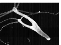
base. We focus especially calcium and lithium. We can show that a dcrease of the calcium concentration in the culturemedium to the intracellular level leads to the formation of branched animals (s. figure). 

This effect is not compensable through application of magnesium or other comparable ions belonging to the same group like strontium or barium. Lithium which interferes with the phosphatidylinositol-cycle is also involved in the pattern forming system of hydra. It is possible to destroy the i-cells in the aminals after application and furthermore bud separation is inhibited. In Hydra vulgaris treatment with LiCl induces the formation of ectopic feet along the body axis. We are searching for the links between phosphorylation and the ions. The foot formation at the bud´s base is used as our testsystem. 


Publications:

Hassel, M., and Berking, S. (1990). Lithium ions interfere with pattern control in Hydra vulgaris. Roux's Arch. Dev. Biol. 198, 382-388.
Hassel, M. und Berking, S. 1988. Nerve cell and nematocyte production in Hydra is deregulated by lithium ions. Roux´s Archiv. Dev. Biol. 197: 471-475.
Pérez, F. (1996). Effects of cantharidin and a phorbol ester on bud formation in Hydra vulgaris. J. Dev. Biol. Suppl. I, 273.
Pérez, F., and Berking, S. (1994). Protein kinase modulators interfere with bud formation in Hydra vulgaris. Roux's Arch. Dev. Biol. 203, 284-289.

Stefanie Zeretzke, Fernando Pérez, Kirsten Velden and Stefan Berking (2002) Ca2+-ions and pattern
      control in Hydra. Int. J. Dev. Biol. 46:705-710 (pdf-file


home

 
Control of spacing of polyps

In Eirene viridula new polyps are formed on adult stolons in a distance of 1,6mm to the last developed polyp. The endogenous substance homarine (N-methypicolinicacid) applied to seawater in concentrations of about 0,1 µM enhanced the distance. The antibiotic sinefungin, a competitor of S-adenosylmethionin (SAM), shortens this distance. It is possible that homarine is used as a donor of a methylgroup in the process of transmethylation for the formation of SAM. Therefore we assume that transmethylation plays an important role in the control of keeping a distance between polyps in a colony. 

Publications:

Berking, S. (1986). Transmethylation and control of pattern formation in Hydrozoa. Differentiation 32, 10-16.


home
Control of polyp polymorphism
 inThecocodium quadratum

Thecocodium quadratum (Werner, Jber. Biol. Anst. Helgoland, 1965) is a colonial hydroid which produces 2 different types of polyps: gastrogonozooids and dactylozooids. The mouthless dactylozooids bear tentacles and catch the prey, which is then taken over and swallowed by the gastrogonozooids which have no tentacles. It is obvious that for a colony to survive both polyps must exist simultaneously arranged in a certain spatial pattern. Our experiments indicate that the formation of polyps in a growing culture is governed by at least 3 principles: (1) short range inhibition between polyps irrespective of their differentiation; (2) long range specific inhibition between gastrogonozooids; and (3) long range supporting influence (lateral help, Meinhardt, H., Models of Biological Pattern Formation, 1982) between gastrogonozooids and dactylozooids.
 

Publications:

Pfeifer, R., and Berking, S. (1995). Control of formation of the two types of polyps in Thecocodium quadratum (Hydrozoa, Cnidaria). Int. J. Dev. Biol. 39, 395-400.


home

 
Thecates

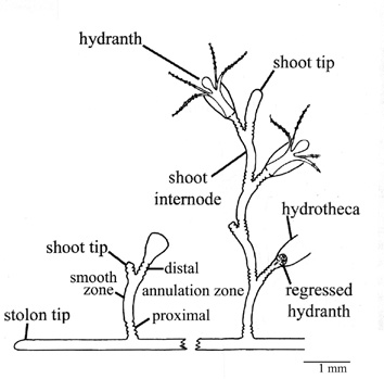
Schematic drawing of a colony of  Laomedea flexuosa

The colonies of thecate hydroids are covered with a chitinous tubelike outer skeleton, the perisarc. The perisarc shows a species-specific pattern of annuli, curvatures, and smooth parts. This pattern is exclusively formed at the growing tips at which the soft perisarc material is expelled by the underlying epithelium. Just behind the apex of the tip, this material hardens. We treated growing cultures of Laomedea flexuosa with substances we suspected would interfere with the hardening of the perisarc (L-cysteine, phenylthiourea) and those we expected would stimulate it (dopamine, N-acetyldopamine). We found that the former caused a widening of and the latter a reduction in the diameter of the perisarc tube. At the same time, the length of the structure elements changed so that the volume remained almost constant. We propose that normal development involves a spatial and temporal regulation of the hardening process. When the hardening occurs close to the apex, the diameter of the tube decreases. When it takes place farther from the apex, the innate tendency of the tip tissue to expand causes a widening of the skeleton tube. An oscillation of the position at which hardening takes place causes the formation of annuli.

Kossevitch IA, Herrmann K, Berking S. (2001) Shaping of Colony Elements in Laomedea flexuosa Hinks (Hydrozoa, Thecaphora) Includes a Temporal and Spatial Control of Skeleton Hardening. Biol Bull. 201(3):417-23.


home